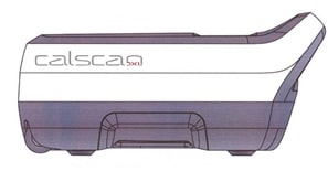
DXL Calscan- a clinically proven diagnostic tool
With over 40 clinical studies published in leading scientific journals around the world, Calscan is a clinically proven tool for any health care center or private physician office. In a hospital environment, Calscan can be used as a stand-alone tool or as an effective complement to an axial DXA device.
What do the experts say?
Prospective hip fracture study of 4,398 females aged 50-99 years, with an average follow-up of 3 years 11 months or 17,200 person years resulting in 130 hip fractures. Conclusion:
“The age-adjusted AUC (area under receiver operating curve) of DXL of calcaneus to predict future hip fractures was 0.84, which is better than that previously reported for DXA of the femoral neck. Of the patients who sustained a hip fracture 78% had a DXL T-score of <-2.5. DXL of calcaneus may therefore be suitable for diagnosing osteoporosis and for predicting fracture risk.”
Brismar TB, Janzky I, Toft L
Vertebral fracture study including 588 subjects, at least one prevalent vertebral fracture identified by X-ray was detected in 160 of them.
“Regarding the identification of subjects with prevalent vertebral fracture, low BMD determined by DXA at the femoral neck or DXL at the calcaneus generally is superior compared to measurements at the lumbar spine.”
“When clinical risk factors were combined with BMD results, the sensitivity and specificity as expressed by AUC (area under receiver operating curves), was identical for DXL calcaneus and iDXA femoral neck at AUC = 0.869. “There was no difference whether BMD was determined at the hip or calcaneus.”
C. Muschitz, H.P. Dimai, R. Kocijian, A. Zendeli, F.Kühne, A. Trubrich, S. Lung, R. Waneck, Heinrich Resch
“DXL Calscan identified correctly significantly more cases of clinical osteoporosis than axial DXA. This was due to the lack of sensitivity of the hi[ results and the falsely elevated scores often found in spinal scans of the elderly.”
Rodionova, SS, Morozov, et al
“We conclude that BMD values obtained with DXA and DXL correlate well and that the DXA and DXL techniques effectively identify the same individuals with low BMD.”
Söderpalm AC, Kullenberg R, Swolin-Eide D
“DXL Calscan provides a more accurate measure of calcaneal BMD than traditional DXA instruments.”
Hakulinen M, Saarakkala S, et al
“We conclude that DXL measurement at the heel bone, using a T-score threshold of -2.5 for classification of osteoporosis, is in concordance with the World Health Organization (WHO) definition of osteoporosis.”
Kullenberg R, Falch J
“Our data showed that DXL Calscan Provides a convenient method of measuring skeletal BMD with some advantages over axial BMD. Calscan diagnostic capacity and relationships with other sites of the skeleton are excellent.”
Martini G, Valenti R, Giovani S, Gennari L, Salvadori S, Galli B, Nuti R
“The Calscan is well suited for use in the management of post-menopausal osteoporosis.”

